Warm Autoimmune Hemolytic Anemia


This patient presented with dyspnea, fatigue, and exercise intolerance. He has stable vital signs, scleral icterus, splenomegaly, but no LAD.

Labs show WBC 5, Hgb 8, PLT 160. CBC previously normal. A DAT (Coombs test) for IgG is positive. The patient has the above peripheral blood smear.


This patient is anemic with scleral icterus which lends itself to a wide differential - one of the most important being hemolysis. The question is where are the blood cells being hemolyzed… one of the most basic things an internist can do to differentiate intravascular vs extravascular hemolysis is to look at the blood cells!

What does this slide show? It does not show any schistocytes which would indicate intravascular hemolysis. It shows spherocytes which is what happens to RBCs when they are opsonized and eventually taken to the spleen to be devoured.

What else shows spherocytes? Hereditary spherocytosis! One way to differentiate these is to get a Coombs test to answer the question, “are there antibodies to these RBCs?”

This patient’s Coombs test is positive. What does that tell you?

  • Warm Agglutinin Disease - IgG - DAT positive for IgG, negative for complement activation

  • Cold Agglutinin Disease = IgM - DAT negative IgG, positive for complement activation

TCA Overdose

This is a fun one. The EKG is consistent with TCA overdose and a pretty nasty one at that. So this patient likely presented with:

So how do we know that 1) this is a TCA overdose and 2) that it is a bad one.

Na Blockade Shifting the DEpolarization to the right

Na Blockade Shifting the DEpolarization to the right

When you look at the EKG you notice that this person is tachycardic, the QRS is wide, and AVR does not look quite normal - that is because there is a Terminal R-Wave - the deflection of the QRS complex is positive at the end. While this is not specific to TCAs and is caused by sodium channel blockade, TCAs are the most common cause. Other examples of Na-blockers include quinidine, mexilitine, phenytoin, lidocaine, and procainamide.

Look to the chart on the right to see where these anti-arrhythmic drugs work. The sodium blockers are going to slow the influx of Na into cells, slowing the action potential - what would you expect to happen to the QRS if the slope of the line at point 0 is not as acute? At the chart on the left you can see that Na-blockade shifts the whole depolarization to the right - prolonging the QRS.

Can you use the QRS to see what people are at risk for? Yes! (You knew that by virtue of me asking…) In TCA overdoses, the QRS width is correlated with toxicity.

  • QRS > 100 ms is predictive of seizures

  • QRS > 160 ms is predictive of ventricular arrhythmias (e.g. VT)

How do you treat this? If you don’t know…maybe time to read that UpToDate Article again.

Vent 101

So, you have correctly identified that a patient needs mechanical ventilation - can’t oxygenate, can’t ventilate, can’t protect their airway, poor anticipated clinical course, or can’t control their secretions - but now what do you do?

  • Select a Mode: Assist-Control vs SIMV vs PSV

  • Select a Target: Volume control, pressure control

  • Set some Variables: rate, TV, Pressure PEEP, FiO2

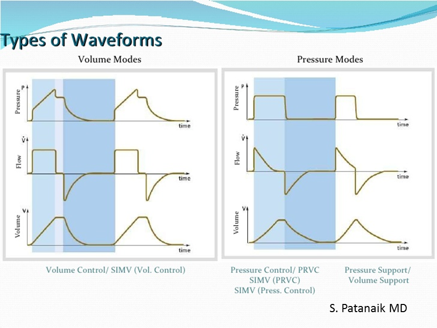
There is often confusion about the mode and the target of the ventilator. The Mode refers to how the ventilator interacts with the patient, whereas the Target is how the ventilator measures and delivers breaths. The Assist-Control mode on a ventilator ensures that all breaths are the same; if the patient triggers the breath it will be assisted to reach the set Target (volume or pressure) or if the patient does not trigger a breath the ventilator will deliver one. In Synchronized Intermittent Mandatory Ventilation, the ventilator will have a set number of mandatory breaths with a target (pressure or volume), but patient triggered breaths will not be strictly “controlled.” These patient triggered breaths may be given some assistance in the form of pressure-support but without a specific target in the vent’s mind. On the right, note the different in the spontaneous breaths between the two charts.

When considering the ventilator settings, think about the Three T’s:

ventwaveforms.png
  • What triggers a breath - patient or time?

  • What is the ventilator targeting - flow or pressure?

  • How does the vent know when to terminate a breath - volume or time?

This breakdown will help you make sense of the flow curves on the vent. Looking to the picture comparing volume and pressure modes, take note at which curve is flat on top - this is the part of the breath that the vent is targeting. In pressure-control, the pressure is set. In volume control, the flow is set - because you can’t measure volume without “flow x time.”

Gene Novikov, MD

Serotonin Syndrome

hocm.gif

Serotonin Syndrome

Serotonin syndrome is often related to iatrogenic causes - medication interactions and intentional self-poisoning - which develops over the course of hours. The classic triad for recognition we all learn is:

  • Mental Status Changes - anxiety, agitation, restlessness, disorientation

  • Autonomic Hyperactivity - diaphoresis, tachycardia, hypothermia, HTN, diarrhea, vomiting

  • Neuromuscular Abnormalities - tremor, muscle rigidity, myoclonus, hyper-reflexia, b/l babinski (all more pronounced in lower extremities)

Diagnosis is made clinically using Hunter Criteria, patient must be taking one serotonergic agent and meet any ONE of the following:

  • Spontaneous clonus

  • Inducible clonus PLUS agitation or diaphoresis

  • Ocular clonus PLUS agitation or diaphoresis

  • Tremor PLUS hyperreflexia

  • Hypertonia PLUS fever PLUS ocular clonus or inducible clonus

The big question is how do you differentiate the other conditions that make people hot and altered?

  • NMS: Also has hyperthermia, AMS, elevated CK/LFTs, and even muscle rigidity - but - NMS is subacute (days to weeks) instead of acute (hours), is more associated with bradyreflexia, and severe rigidity (buzz word: lead pipe rigidity).

  • Anticholinergic Toxicity: Also has agitation, AMS, hyperthermia - but - take care to go a good MSK exam and note the reflexes and tone which should be normal in the anticholinergic toxidrome

  • Malignant Hyperthermia: Also has rigidity, tachycardia, hyperthermia, and acidosis - but - the clinical situation is different as MH is usually related to halogenated volatile anesthetics and depolarizing muscle relaxers.

Treatment is largely supportive care, but also keep these key steps in mind (they’ll be on boards)

  1. Discontinue offending drugs

  2. Sedate with BZD

  3. If that does not help -> Cyproheptadine

    1. When administered as an antidote for serotonin syndrome, an initial dose of 12 mg is recommended, followed by 2 mg every two hours until clinical response is seen. Cyproheptadine is only available in an oral form! You will likely have to place an NG to administer this medication

Gene Novikov, MD

HOCM

Hypertrophic Cardiomyopathy (HOCM)

HOCM is an autosomal dominant heritable disorder that results in increased LV wall thickening, abnormal LV filling with a dynamic LV outflow tract obstruction and LV diastolic dysfunction due to stiff/hypertrophied LV. This clinically presents with dyspnea, presyncope/syncope, or sudden cardiac death during exercise. Patients with have a systolic murmur best heard at the LLSB that increases with Valsalva (decreases venous return) and decreases with squatting/leg raise (increase venous return). ECG will commonly demonstrate increased QRS voltage, LA enlargement, LVH, pathologic Q waves (dagger-like) in the lateral + inferior leads, and repolarization abnormalities.

Diagnosis is established by TTE which shows dynamic LVOT obstruction. After TTE diagnosis, patient needs evaluation for arrhythmia by 24-hour ECG monitoring or functional hypotension by exercise stress testing which are predictors of sudden cardiac death (SCD).

Patients may require primary prophylaxis of SCD with ICD placement if:

  • First degree relative with SCD

  • LV wall thickness >30 mm

  • Syncope

  • Nonsystained VT

  • Abnormal BP during treadmill exercise

Patients should be advised to avoid high-intensity sports, dehydration (diuretics) and vasodilation (heat, nitrates, phosphodiasterase type 5 inhibitors, dihydropyridine calcium channel blockers). Medical therapy includes beta-blocker (avoid carvedilol, labetalol which have vasodilatory component) and non-dihydropyridine calcium channel blockers (verapamil, diltiazem).

Septal myectomy or septal ablation may be considered in patients with :

  • mod-severe obstruction despite maximal medical therapy + LVOT gradient >50 mmHg

  • recurrent syncope

Surveillance is indicated in patient and first degree relatives. Patients should undergo 24-hour ECG monitoring every 1-2 years. Family members should receive genetic counseling, with ECG and TTE testing every 5 years.

- Natalie Hood, MD