Spine Sign

hocuspocus.gif

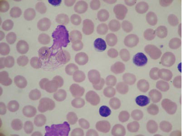
A 50 YEAR OLD PATIENT WITH ALCOHOL ABUSE PRESENTS WITH SEVERE EPIGASTRIC ABDOMINAL PAIN AND DYSPNEA. YOU OBTAIN THE BEDSIDE US CAPTURED ABOVE. WHAT SINGLE LABORATORY TEST WILL HELP CONSOLIDATE THIS PATIENT'S PRESENTATION?

Spine Sign

We had a fantastic faculty session with Dr. Jessica Baez on Point of Care Ultrasound for the lungs. Check that out here if you missed it! We’re continuing on the POCUS train with review of “spine sign” in pleural effusion.

spine-sign-present.jpg

Positive Spine Sign is a clear view of several thoracic vertebrae through the effusion. If you look closely at the ultrasound above, you can see the outline of several vertebra. It is 92% sensitive and ~93% specific for presence of a pleural effusion.

spine-sign-absent.jpg

Negative Spine Sign: Let’s compare with normal lung showing a negative Spine sign. In normal lung, the air within the lungs should obstruct your view of the thoracic vertebra.


In the patient in our case with alcohol abuse presenting with severe epigastric abdominal pain and dyspnea. His bedside US demonstrates a pleural effusion. In the context of alcohol abuse with epigastric pain, my differential includes gastritis, PUD, pancreatitis, Mallory-Weiss Tea, Boerhaave, acute hepatitis; less likely: ACS, pulmonary embolism, aortic ulcer/dissection. With the evidence of pleural effusion based on spine sign, I begin thinking about how any of these differential diagnoses can lead to an effusion. My differential narrows to esophageal perforation (if clinical history of significant retching) versus acute pancreatitis with pleural effusion. A single laboratory test performed on pleural fluid that could consolidate the abdominal pain and the effusion would be a pleural fluid amylase level. Pleural fluid amylase >ULN for serum amylase, or a pleural fluid : serum amylase ratio > 1.0, is indicative of pancreatitis or esophageal rupture. In the case above, the patient has an pancreatic pleural effusion with ascites.

Further references: Extension of Thoracic Spine Sign

~Natalie Hood, MD

Glomitis

Screen+Shot+2020-04-24+at+1.40.50+PM.png

Glomerulonephritis

The impressive Michelle Sandoval created a review module on Glomerular Disease that you should definitely check out.

This urinalysis shows an RBC cast and suggests glomerular disease / inflammation at the level of the glomerulus. Erythrocytes may be distinct or incorporated into a homogenous mass. The most characteristic feature of a RBC cast is its orange-red appearance.

This is a great time to review a little about how we think about hematuria. The first question is always: Is it real? So confirm via repeat test and verification that bleeding is from urinary tract rather than mucosa trauma or vaginal source.

After you’ve confirmed that it really is hematuria, your next goal should be identifying the source as upper versus lower tract. This is where the urine sediment comes into play. For lower tract (i.e. NONglomerular) bleeding, you will see frank clot or normal looking erythrocytes. For upper tract (i.e. glomerular) bleeding, you will see RBC casts and dysmorphic RBCs. Once you have differentiated upper from lower tract, you can now target your next step to visualize the source. Glomerular bleeding warrants renal ultrasound and further work up of etiology whereas lower urinary tract bleeding warrants cystoscopy for direct visualization.

image-asset.png

TB

ctcavitation.png

A patient with the above cavitary lesion is diagnosed with active TB. He wants to know when he will be considered non-contagious?

TB Treatment

There are 3 criteria to be met before a patient with active TB is deemed “noncontagious”:

1.) adequate treatment for at least 2 weeks (with rifampin, isoniazid, pyrazinamide, and ethambutol- RIPE)

2.) improvement of symptoms

3.) three consecutive negative sputum smears (smears collected at 8- to 24-hour apart intervals with one smear as an early morning collection).

Vitamin K Deficiency

You are rounding on your patient, a 64 y/o M with C.Diff after treatment with antibiotics for CAP. He has had poor PO 2/2 nausea. He is on PO Vanc, Unasyn, and SQH. In the am, you are notified of prolonged bleeding thing from his PIVs, bruising, and see the above on exam. Labs show:

APTT WNL
PT Prolonged
HGB 12 (PRIOR 12.8)
PLT 190
LFT AND Factor VIII, V are WNL

What is the most likely etiology of your patient's bleeding? How will you intervene?


Vitamin K Deficiency

Let’s walk through this one! Last week Dr. Espinoza gave a great Fellow’s Friday on Bleeding Disorders. Refer back to that video under Distance Learning tab if you missed it.

Any patient with prolonged bleeding merits laboratory work up to evaluate their hemostasis with a prothrombin time (PT) and an activated partial thromboplastin time (aPTT). As you work through this bleeding patient’s coagulation studies, utilize the coagulation cascade to guide you.

There are three arms of the coagulation cascade: the intrinsic (aPTT; factors XII, XI, IX, VIII), extrinsic (PT; factor VII), and the common pathway (aPTT/PT; additional factors X, V, II).

Recall that Heparin use inhibits the intrinsic pathway and Coumadin use inhibits the extrinsic pathway. Recall that Vitamin K dependent factors are FX, IX, VII, II, protein C & S (bonus mnemonic: “1972 was the disco era”).

Our patient had a normal aPTT, prolonged PT, normal PLT count, and factors VIII and V were wnl.

The normal aPTT makes a deficiency / inhibitor along the intrinsic pathway unlikely – ruling out common etiologies like Hemophilia A (FVIII) or Hemophilia B (IX), vWD (VIII), those factor inhibitors, or heparin use.  If the common pathway was involved, we would expect both aPTT and PT to be abnormal so we also can rule out problems with FX, V, and II.

The PT was prolonged so we should focus more on this side of the cascade. Our differential can include FVII deficiency/inhibition (Coumadin use, vitamin K deficiency), DIC, or liver disease.

For DIC or liver disease, we would expect the PLT count to be low and for factor VII to be low. If both of those values had been low, we could’ve then used factor VIII to differentiate between DIC or liver disease. In DIC, the coagulation cascade is activated and consumed meaning that all factors we test for should be low. In liver disease, all factors that are synthesized by the liver should be low. Factor VIII is a helpful test though because it is not only produced by the liver but also synthesized by endothelial cells. FVIII will therefore be normal in liver failure (or even elevated).

Looking at the clinical context: Our patient is not on Coumadin. He has been hospitalized with pneumonia and c.diff, on multiple antibiotics, and has had poor nutrition through all of this. Our leading diagnosis is therefore vitamin K deficiency.

Although vitamin K is found in many foods (think leafy greens), actually most of your vitamin K is derived from colonic bacteria. Low dietary intake alone is rarely the cause for severe deficiency. His recent antibiotics likely wiped out his normal bacteria and his active diarrhea likely decreased is absorption from eating. Add in the poor appetite… And presto.

So why did the PT prolong, but the aPTT was normal? The PT is most sensitive and earliest sign of a decrease in vitamin-K dependent factors and PT will be prolonged out of proportion to the aPTT which will be normal or only mildly elevated. In severe vitamin K deficiency both PT and PTT will prolong.

Treatment options: IV vitamin K 10 mg is sufficient to restore normal clotting factor levels within <10 hours. If a patient has ongoing bleeding or need for immediate correction before an invasive procedure, then FFP or PCC is your answer. In an asymptomatic patients without active bleeding or high risk, enteric vitamin K is sufficient.

Takotsubo's

tak.gif

The above TTE was a apical 4 chamber view that showed apical ballooning consistent with Tatoksubo’s cardiomyopathy! Sometimes it’s hard to tell what the ventricular walls are doing to the untrained generalists eyes. One cardiologist’s tip was to place your finger in the middle of the chamber and isolate each walls movement in reference to your finger. When you do that, you can definitely see that the walls in the apex are not moving at all and are ballooned out. See the fig to the right.

Takotsubo’s is an important item to keep on your differential when seeing patients with chest pain as 1-2% of troponin-positive suspected ACS patients have this stress induced cardiomyopathy.

Can you identify the view on the TTE above? Can you identify which chambers are which? Did this look abnormal to you? If the answer is “no” to any of these - check out these lectures on echos!

Peptic Esophageal Stricture

peptic.jpg

Peptic Esophageal Stricture

Peptic Esophageal Strictures are a subset of benign esophageal strictures, so let’s review different etiologies: peptic, radiotherapy-induced and caustic injuries, Schatzki ring, eosinophilic esophagitis (EoE), and strictures after surgical resection (anastomotic) or endoscopic resection.

Peptic strictures are a result of the healing process of ulcerative esophagitis. Collagen is deposited during this phase and, with time, the collagen fibers contract, narrowing the esophageal lumen. These strictures are usually short in length and contiguous with the gastroesophageal junction. Patients may have solid food dysphagia and episodic food impaction. The management of benign esophageal strictures involves dilation combined with acid-suppressive therapy with a proton pump inhibitor to prevent the recurrence of strictures once they have been adequately dilated.

SVT w/ Aberrancy

SVT vs VT

This is the age old question when you’re in a rapid response with a wide-complex tachyarrhythmia - is this VT or is this SVT? There are a few helpful criteria (Brugada’s Criteria) that will help you differentiate these. We have made a module for you to practice identifying these - the answers are there but the explanations will be posted next week. See if you can figure out what is SVT and what is VT.

VT vs SVT Module

SJS

SJS3.jpg

So the clinical picture may be difficult to decipher without a history or full skin exam, but let’s use some of the answers we got to discuss the skin findings above.

One of the answers submitted said it Kaposi Sarcoma, see to the left. The classic presentation of Kaposi’s is multiple violaceous papules and plaques. Make note of the COLORING of the lesions. In contrast, the trivia rash is made up of atypical target lesions. Take a close look at the rash and you will see that there is a dark center with a lighter erythematous border, 2 areas of color. Typical target lesions have THREE areas, often seen in Erythema Multiforme and Lyme Disease.

On the right was another submission - Purpura Fulminans. Again this rash is made up of retiform purpura with branched or angular purpuric lesions. Made note of the SHAPE of the lesions, the picture on the right shows purpura fulminans. Again, our rash had atypical target lesions with the start of some bullae formation.

The trivia rash is of early SJS, which can be characterized by atypical target lesions. Make sure to check their oral mucosa and take a good medication history.

As I was preparing for AHD: Derm, I was curious about the pathophysiology of DRESS and SJS - they are both drug reactions, so why are they so different? Looking into the pathophysiology, they are both the result of a medication activating t-cells via MHC pathway. In SJS, this leads to Killer T-Cells attacking Keratinocytes - causing a rash within days of exposure and causing desquamation. SJS is not typically associated with significant systemic issues. DRESS, however, is thought to be a result of viral reactivation after t-cell activation. This could be why the DRESS is often more delayed to weeks after exposure, the rash is morbiliform, it resembles viral exanthems, and is associated with -itis’s of different organs.

Pulmonary Alveolar Proteinosis

A 28 yo HIV-negative man presents with several month history of progressively worsening dyspnea and nonproductive cough.

His oxygen saturation was 75% on room air. On exam , he has digital clubbing. Lungs were CTAB.

Computed tomography of the chest (above) revealed diffuse ground-glass opacities with interlobular and intralobular septal thickening — a pattern described as crazy paving pattern. Bronchoscopy with lavage revealed milky-appearing fluid, with macrophages. BAL testing was positive on periodic acid–Schiff staining.

Pulmonary Alveolar Proteinosis

Pulmonary alveolar proteinosis (PAP) is a lung disease characterized by an abnormal intra-alveolar accumulation of surfactant-derived lipoproteinaceous material with a fairly vague and non-specific clinical presentation: dyspnea or a minimally-productive cough. It can be divided into 3 main categories: autoimmune, secondary, and congenital (least interesting…). The most common is the autoimmune which is due to an IgG to GM-CSF proteins. The next most common is secondary - or in response to an inflammatory insult: hematologic malignancies, inhalational disease, HIV, immunosuppression.

Cardiac Tamponade

This TTE image is a parasternal long axis view. To obtain a parasternal long axis view, place phase array transducer at the left sternal border in the left 3rd-4th intercostal space with the probe indicator directed towards the patient’s right shoulder.

In this image, you have a large circumferential pericardial effusion causing dynamic diastolic collapse of the RA/RV. This is cardiac tamponade.

TTE+image.png

We can differentiate pleural effusions from pericardial effusions by looking for the location of the anechoic fluid relative to the aorta.

  • When the fluid is between the aorta and the LA/LV it is pericardial.

  • When the fluid extends behind the aorta (closer to the bottom of the screen), it is pleural.

Normally, there is ~10mL of pericardial fluid to lubricate the space between the heart and the pericardial sac. Cardiac tamponade is not a volume-dependent phenomenon, but can occur at low volumes accumulating over a short amount of time. Therefore, all patients with pericardial effusions should be closely evaluated.

download2.jpg

Clinical cardiac tamponade is associated with Beck’s triad (hypotension, muffled heart sounds, and JVD); however, this is fairly uncommon in practice. Echocardiographic cardiac tamponade is TTE features of tamponade physiology in the asymptomatic patient.

In the hypotensive fluid with cardiac tamponade, bolus fluids and emergent pericardiocentesis is recommended. Pericardial window can be performed, but if the underlying etiology off the effusion is not addressed, the effusion will recur as the pericardial sac scars down after window. Definitive treatment is targeting the underlying cause.

Screen+Shot+2020-03-06+at+1.31.12+PM.png

Perioperative Medicine

Perioperative-and-Surgical-Nursing.jpg

Let’s talk perioperative medicine! Which patients should get a pre-operative ECG?

As we learned this week in AHD, perioperative medicine is incredibly variable. Our expert Reza explains:

The 2014 ACC/AHA guideline regarding this issue uses vague language. Here is what it says:

Preoperative resting 12-lead ECG is reasonable for patients with known coronary heart disease, significant arrhythmia, peripheral arterial disease, cerebrovascular disease, or other significant structural heart disease, except for those undergoing low-risk surgery.​

​Preoperative resting 12-lead ECG may be considered for asymptomatic patients without known coronary heart disease, except for those undergoing low-risk surgery.​

Routine preoperative resting 12-lead ECG is not useful for asymptomatic patients undergoing low-risk surgical procedures.​

What does this mean practically?

First of all, if a patient is going for minor surgery such as dermatological procedure or cataract surgery, probably don't need to worry about getting an ECG. For patients who are at elevated risk for major adverse cardiac event (elevated risk = > 1%), we should probably have a recent ECG (within 1-3 months) in the chart prior to proceeding with surgery. ​

Reza Ghoorkhanian, MD

Quant Gold Interpretation

download.png

What is the interpretation of the below test result and what would be your next steps?

The Nil = this is your negative control tube. This should not get stimulated to produce interferon. If it is positive, this suggests there is something wrong with the test.

The Mitogen = this is your positive control tube. This should get stimulated to produce interferon. If it is negative, this suggests there is something wrong with the immune response (anergy, immune suppressed).

The TB1, TB2 = these are TB peptides. If the patient has been exposed to TB, this should generate interferon.

In the test tubes above, the Nil is very positive rendering this an inaccurate test. There is too much background inferferon production which has no relation to TB. This immune system is on fire and we are actually looking at a patient with HLH.

Link to Dr. Luckett’s MGR slides + audio

PJP Pneumonia

This 28 yo M with pmh of severe rheumatoid arthritis presents with shortness of breath and cough for 3 months. He has been treated as outpatient for presumed community acquired pneumonia but has failed to improve. He presents with worsening SOB.

On arrival, T 99.2, HR 108, BP 113/50, RR 20, SpO2 89%.

Exam: slightly increased WOB with tachypnea. Diffuse crackles. Otherwise unremarkable. No LAD, meningismus.

Labs: WBC 12.5, Hgb 11, PLT 309; BMP remarkable for HCO3 21, normal renal function. LDH 413. ABG 7.45/34/60/21 (A-a gradient 47)

Home meds: Methotrexate, abatacept, prednisone, percocet.

PJP Pneumonia

This is an immunocompromised patient with acute hypoxic respiratory failure due, subacute in onset, with a reticulonodular pattern on their CXR - makes you worry about PJP Pneumonia! But since this patient does not have HIV - why are they at risk? While there are no published guidelines on PJP ppx for most immunocompromised patients (other than HCT patients), a meta-analysis showed that patients with a greater than 6% chance of of PJP warrant ppx. For our purposes, the highest yield population in this group is the following: patients receiving a glucocorticoid dose >20mg/d for one month or longer who also have another cause of immunocompromise (hematologic malignancy or a second immunosuppressant medication). While we do not know the doses of the prednisone in this patient, the clinical picture suggests PJP in the setting of multiple immunosuppressants.

Interestingly, LDH is often used as a biomarker in PJP. This is only sensitive in the HIV-infected population! In the HIV-uninfected population, there are so many other causes of immunodeficiency that concurrently increase your LDH that it becomes significantly less sensitive.

The question asks what to start for treatment and treatment dosed bactrim is the correct answer - but this patient also warrants adjunctive glucocorticoids. This is based on the degree of respiratory compromise as seen on the ABG and A-a (Alveolar-arterial) gradient. A PO2 < 70 and A-a > 35 requires steroids. Steroids lessen the inflammatory response from the dying PJPs and improves morbidity and mortality.

Smudge Cells in Chronic Lymphocytic Leukemia

9a8631b5-460a-4fd7-a8c3-5be7b85c901b.png

Smudge Cells in Chronic Lymphocytic Leukemia

Chronic lymphocytic leukemia/small lymphocytic lymphoma (CLL/SLL) is a mature B cell neoplasm characterized by a progressive accumulation of monoclonal B lymphocytes. The lymphocytes are predominantly small and mature appearing, and due to increased fragility they may form “smudge cells” on the peripheral smear (as pictured here). The frequency of smudge cells can have prognostic implications in CLL. A study out of Mayo Clinic found that >30% smudge cells is associated with more aggressive form of CLL. Smudge cells can also be due to infectious causes or simply due to an old blood sample.

CLL is recognized based on increased absolute lymphocyte count on CBC. Patients can describe B cell symptoms and can have hepatosplenomegaly and lymphadenopathy. Patients are often immunosuppressed due to hypogammaglobulinemia and may present with recurrent bacterial infections.

One of the significant complications of CLL is leukostasis. While leukostasis is more common to AML & CML with WBC >100x10^9/L and symptoms of impaired tissue perfusion, it can occur in CLL when WBC >400x10^9/L. Patients can present with pulmonary (dyspnea, hypoxia, falsely reduced PaO2), neurologic (HA, vision changes, AMS), and fever. This is a medical emergency! 20-40% of patients ultimately die within 1 week of presentation. Treatment: aggressive hydration and cytoreduction (via induction chemotherapy, hydroxyurea, versus leukapheresis).

Post-MI Pericarditis

ITE Round Up: Post MI Pericarditis

peri.png

You are managing a patient in CVICU who is 2 days s/p PCI to LAD after presenting with STEMI. He is now describing chest pain that improves with leaning forward. You think you hear a cardiac rub. You have obtained the above ECG. How will you treat this patient?

We often think about STEMIs as a single vessel problem, we describe the pathology using terms like “target lesion” but there is a significant systemic inflammatory component to an MI that we overlook. When thinking about the complications of an MI we can break it down into two categories - mechanical complications (ruptures of all sorts depending on the amount of tissue necrosis) and inflammatory/immunologic (pericarditidis of all sorts).

First things first, how do we diagnose pericarditis?

  • Chest pain – Typically sharp, pleuritic, and centrally located; the pain is improved by sitting up and leaning forward.

  • Pericardial friction rub – A superficial scratchy or squeaking sound best heard with the diaphragm of the stethoscope over the left sternal border; the rub is frequently intermittent and may be difficult to appreciate, but is diagnostic when present.

  • ECG changes – New widespread ST elevation and/or PR depression which extend beyond a typical anatomic regional boundary.

  • Pericardial effusion – Most effusions are small and not hemodynamically significant

Next. Is this Dresslers Syndrome or Peri-Infarction Pericarditis (PIP)? You can use timeline to help with this. Dressler’s is a delayed response after several weeks. How do we treat these?

PIP is usually a self-limited process related to inflammation from an MI. If symptoms are prolonged past a week, ASA 650mg q6-8h is recommended. There is no role in colchicine or glucocorticoids.

Dressler’s is treated similarly to acute pericarditis - NSAIDs (ASA or Ibu), colchicine is an option, glucocorticoids.

Gene Novikov, MD

Warm Autoimmune Hemolytic Anemia


This patient presented with dyspnea, fatigue, and exercise intolerance. He has stable vital signs, scleral icterus, splenomegaly, but no LAD.

Labs show WBC 5, Hgb 8, PLT 160. CBC previously normal. A DAT (Coombs test) for IgG is positive. The patient has the above peripheral blood smear.


This patient is anemic with scleral icterus which lends itself to a wide differential - one of the most important being hemolysis. The question is where are the blood cells being hemolyzed… one of the most basic things an internist can do to differentiate intravascular vs extravascular hemolysis is to look at the blood cells!

What does this slide show? It does not show any schistocytes which would indicate intravascular hemolysis. It shows spherocytes which is what happens to RBCs when they are opsonized and eventually taken to the spleen to be devoured.

What else shows spherocytes? Hereditary spherocytosis! One way to differentiate these is to get a Coombs test to answer the question, “are there antibodies to these RBCs?”

This patient’s Coombs test is positive. What does that tell you?

  • Warm Agglutinin Disease - IgG - DAT positive for IgG, negative for complement activation

  • Cold Agglutinin Disease = IgM - DAT negative IgG, positive for complement activation

Vent 101

So, you have correctly identified that a patient needs mechanical ventilation - can’t oxygenate, can’t ventilate, can’t protect their airway, poor anticipated clinical course, or can’t control their secretions - but now what do you do?

  • Select a Mode: Assist-Control vs SIMV vs PSV

  • Select a Target: Volume control, pressure control

  • Set some Variables: rate, TV, Pressure PEEP, FiO2

There is often confusion about the mode and the target of the ventilator. The Mode refers to how the ventilator interacts with the patient, whereas the Target is how the ventilator measures and delivers breaths. The Assist-Control mode on a ventilator ensures that all breaths are the same; if the patient triggers the breath it will be assisted to reach the set Target (volume or pressure) or if the patient does not trigger a breath the ventilator will deliver one. In Synchronized Intermittent Mandatory Ventilation, the ventilator will have a set number of mandatory breaths with a target (pressure or volume), but patient triggered breaths will not be strictly “controlled.” These patient triggered breaths may be given some assistance in the form of pressure-support but without a specific target in the vent’s mind. On the right, note the different in the spontaneous breaths between the two charts.

When considering the ventilator settings, think about the Three T’s:

ventwaveforms.png
  • What triggers a breath - patient or time?

  • What is the ventilator targeting - flow or pressure?

  • How does the vent know when to terminate a breath - volume or time?

This breakdown will help you make sense of the flow curves on the vent. Looking to the picture comparing volume and pressure modes, take note at which curve is flat on top - this is the part of the breath that the vent is targeting. In pressure-control, the pressure is set. In volume control, the flow is set - because you can’t measure volume without “flow x time.”

Gene Novikov, MD

Serotonin Syndrome

hocm.gif

Serotonin Syndrome

Serotonin syndrome is often related to iatrogenic causes - medication interactions and intentional self-poisoning - which develops over the course of hours. The classic triad for recognition we all learn is:

  • Mental Status Changes - anxiety, agitation, restlessness, disorientation

  • Autonomic Hyperactivity - diaphoresis, tachycardia, hypothermia, HTN, diarrhea, vomiting

  • Neuromuscular Abnormalities - tremor, muscle rigidity, myoclonus, hyper-reflexia, b/l babinski (all more pronounced in lower extremities)

Diagnosis is made clinically using Hunter Criteria, patient must be taking one serotonergic agent and meet any ONE of the following:

  • Spontaneous clonus

  • Inducible clonus PLUS agitation or diaphoresis

  • Ocular clonus PLUS agitation or diaphoresis

  • Tremor PLUS hyperreflexia

  • Hypertonia PLUS fever PLUS ocular clonus or inducible clonus

The big question is how do you differentiate the other conditions that make people hot and altered?

  • NMS: Also has hyperthermia, AMS, elevated CK/LFTs, and even muscle rigidity - but - NMS is subacute (days to weeks) instead of acute (hours), is more associated with bradyreflexia, and severe rigidity (buzz word: lead pipe rigidity).

  • Anticholinergic Toxicity: Also has agitation, AMS, hyperthermia - but - take care to go a good MSK exam and note the reflexes and tone which should be normal in the anticholinergic toxidrome

  • Malignant Hyperthermia: Also has rigidity, tachycardia, hyperthermia, and acidosis - but - the clinical situation is different as MH is usually related to halogenated volatile anesthetics and depolarizing muscle relaxers.

Treatment is largely supportive care, but also keep these key steps in mind (they’ll be on boards)

  1. Discontinue offending drugs

  2. Sedate with BZD

  3. If that does not help -> Cyproheptadine

    1. When administered as an antidote for serotonin syndrome, an initial dose of 12 mg is recommended, followed by 2 mg every two hours until clinical response is seen. Cyproheptadine is only available in an oral form! You will likely have to place an NG to administer this medication

Gene Novikov, MD

HOCM

Hypertrophic Cardiomyopathy (HOCM)

HOCM is an autosomal dominant heritable disorder that results in increased LV wall thickening, abnormal LV filling with a dynamic LV outflow tract obstruction and LV diastolic dysfunction due to stiff/hypertrophied LV. This clinically presents with dyspnea, presyncope/syncope, or sudden cardiac death during exercise. Patients with have a systolic murmur best heard at the LLSB that increases with Valsalva (decreases venous return) and decreases with squatting/leg raise (increase venous return). ECG will commonly demonstrate increased QRS voltage, LA enlargement, LVH, pathologic Q waves (dagger-like) in the lateral + inferior leads, and repolarization abnormalities.

Diagnosis is established by TTE which shows dynamic LVOT obstruction. After TTE diagnosis, patient needs evaluation for arrhythmia by 24-hour ECG monitoring or functional hypotension by exercise stress testing which are predictors of sudden cardiac death (SCD).

Patients may require primary prophylaxis of SCD with ICD placement if:

  • First degree relative with SCD

  • LV wall thickness >30 mm

  • Syncope

  • Nonsystained VT

  • Abnormal BP during treadmill exercise

Patients should be advised to avoid high-intensity sports, dehydration (diuretics) and vasodilation (heat, nitrates, phosphodiasterase type 5 inhibitors, dihydropyridine calcium channel blockers). Medical therapy includes beta-blocker (avoid carvedilol, labetalol which have vasodilatory component) and non-dihydropyridine calcium channel blockers (verapamil, diltiazem).

Septal myectomy or septal ablation may be considered in patients with :

  • mod-severe obstruction despite maximal medical therapy + LVOT gradient >50 mmHg

  • recurrent syncope

Surveillance is indicated in patient and first degree relatives. Patients should undergo 24-hour ECG monitoring every 1-2 years. Family members should receive genetic counseling, with ECG and TTE testing every 5 years.

- Natalie Hood, MD